Please consult with your health-care provider before making any dietary changes based on any of the information provided here.
If you live in Quebec and do not have a family doctor, you can [ sign up here ].
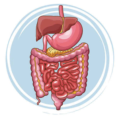
Anatomy and Physiology

Anatomy and Physiology is a dynamic textbook for the two-semester human anatomy and physiology course for life science and allied health majors. The book is organized by body system and covers standard scope and sequence requirements.
More InfoHuman Nutrition

This textbook serves as an introduction to nutrition. The book covers basic concepts in human nutrition, key information about essential nutrients, basic nutritional assessment, and nutrition across the lifespan.
More InfoPrinciples of Nutrition

A great textbook to get started on a journey into the world of health and nutrition. The first focus will be to demonstrate that nutritional science is an evolving field of study, continually being updated and supported by research, studies, and trials.
More InfoMicrobiology

Microbiology covers the scope and sequence requirements for a single-semester microbiology course for non-majors. The book presents the core concepts of microbiology with a focus on applications for careers in allied health. The pedagogical features of the text make the material interesting and accessible while maintaining the career-application focus and scientific rigor inherent in the subject matter.
More InfoChemistry

Chemistry 2e is designed to meet the scope and sequence requirements of the two-semester general chemistry course. The textbook provides an important opportunity for students to learn the core concepts of chemistry and understand how those concepts apply to their lives and the world around them. The book also includes a number of innovative features, including interactive exercises and real-world applications.
More InfoBiology

Biology 2e is designed to cover the scope and sequence requirements of a typical two-semester biology course for science majors. The text provides comprehensive coverage of foundational research and core biology concepts through an evolutionary lens. Biology includes rich features that engage students in scientific inquiry, highlight careers in the biological sciences, and offer everyday applications.
More InfoChapter Objectives
After studying dris chapter, you will be able to::
- List and describe the functional anatomy of the organs and accessory organs of the digestive system
- Discuss the processes and control of ingestion, propulsion, mechanical digestion, chemical digestion, absorption, and defecation
- Discuss the roles of the liver, pancreas, and gallbladder indigestion
- Compare andconttast the digestion of the three macronutrients
The digestive system is continually at work, yet people seldom appreciate the complex tasks it performs ina choreographed biologic symphony.Consider what happens when you eat an apple.Of course, you enjoy the apple’s taste as you chew it, but inthe hours that follow,unless something goes amiss and you get a stomachache, you don’t notice that your digestive system is working. You may be taking a walk or studying or sleeping, having forgotten all about the apple, but your stomach and intestines are busy digesting it and absorbing its vitamins and other nutrients.

Figure 23.1 Eating Apples Eating may be one of the simple pleasures in life,but digesting even one apple requires the coordinated work of many organs.
By the time any waste material is excreted, the body has appropriated all it can use from the apple. Inshort, whether you pay attention or not, the organs of the digestive system perform their specific functions, allowing you to use the food you eat to keep you going. This chapter examines the structure and functions of these organs, and explores the mechanics and chemistry of the digestive processes.

Overview of the Digestive System
By the end of this section, you will be able to: :
- Identify the organs of the alimentary canal from proximal to distal, and briefly state their function
- Identify the accessory digestive organs and briefly state their function
- Describe the four fundamental tissue layers of the alimentary canal
- Contrast the contributions of the enteric and autonomic nervous systems to digestive system functioning
- Explain how the peritoneum anchors the digestive organs
The function of the digestive system is to break down the foods you eat, release their nutrients, and absorb those nutrients into the body. Although the small intestine is the workhorse of the system, where the majority of digestion occurs, and where most of the released nutrients are absorbed into the blood or lymph, each of the digestive system organs makes a vital contribution to this process (Figure 23.2). As is the case with all body systems, the digestive system does not work in isolation; it functions cooperatively with the other systems of the body. Consider for example, the interrelationship between the digestive and cardiovascular systems.
Arteries supply the digestive organs with oxygen and processed nutrients, and veins drain the digestive tract These intestinal veins, constituting the hepatic portal system, are unique; they do not return blood directly to the heart. Rather, this blood is diverted to the liver where its nutrients are off-loaded for processing before blood completes its circuit back to the heart At the same time, the digestive system provides nutrients to the heart muscle and vascular tissue to support their functioning. The interrelationship of the digestive and endocrine systems is also critical. Hormones secreted by several endocrine glands, as well as endocrine cells of the pancreas, the stomach, and the small intestine, contribute to the control of digestion and nutrient metabolism. In turn, the digestive system provides the nutrients to fuel endocrine function. Table 23.1 gives a quick glimpse at how these other systems contribute to the functioning of the digestive system.

Figure 23.2 Components of the Digestive System All digestive organs play integral roles in the life-sustaining process of digestion.
Contribution of Other Body Systems to the Digestive System |
|
| Body system | Benefits received by the digestive system |
| Cardiovascular | Blood supplies digestive organs with oxygen and processed nutrients |
| Endocrine | Endocrine hormones help regulate secretion in digestive glands and accessory organs |
| lntegumentary | Skin helps protect digestive organs and synthesizes vitamin D for calcium absorption |
| Lymphatic | Mucosa-associated lymphoid tissue and other lymphatic tissue defend against entry of pathogens; lacteals absorb lipids; and lymphatic vessels transport lipids to bloodstream |
| Muscular | Skeletal muscles support and protect abdominal organs |
| Nervous | Sensory and motor neurons help regulate secretions and muscle contractions in the digestive tract |
| Respiratory | Respiratory organs provide oxygen and remove carbon dioxide |
| Skeletal | Bones help protect and support digestive organs |
| Urinary | Kidneys convert vitamin D into its active form, allowing calcium absorption in the small intestine |
| Tableau 23.1 | |

The easiest way to understand the digestive system is to divide its organs into two main categories. The first group is the organs that make up the alimentary canal. Accessory digestive organs comprise the second group and are critical for orchestrating the breakdown of food and the assimilation of its nutrients into the body. Accessory digestive organs, despite their name, are critical to the function of the digestive system.
Alimentary canal Organs
Also called the gastrointestinal (GI) tract or gut, the alimentary canal (aliment- = “to nourish”) is a one-way tube about 7.62 meters (25 feet) in length during life and closer to 10.67 meters (35 feet) in length when measured after death, once smooth muscle tone is lost. The main function of the organs of the alimentary canal is to nourish the body. This tube begins at the mouth and terminates at the anus. Between those two points, the canal is modified as the pharynx, esophagus, stomach, and small and large intestines to fit the functional needs of the body. Both the mouth and anus are open to the external environment; thus, food and wastes within the alimentary canal are technically considered to be outside the body. Only through the process of absorption do the nutrients in food enter into and nourish the body’s “inner space.”
Accessory Structures
Each accessory digestive organ aids in the breakdown of food (Figure 23.3). Within the mouth, the teeth and tongue begin mechanical digestion, whereas the salivary glands begin chemical digestion. Once food products enter the small intestine, the gallbladder, liver, and pancreas release secretions—such as bile and enzymes—-essential for digestion to continue. Together, these are called accessory organs because they sprout from the lining cells of the developing gut (mucosa) and augment its function; indeed, you could not live without their vital contributions, and many significant diseases result from their malfunction. Even after development is complete, they maintain a connection to the gut by way of ducts.
Histology of the Alimentary Canal
Throughout its length, the alimentary tract is composed of the same four tissue layers; the details of their structural arrangements vary to fit their specific functions. Starting from the lumen and moving outwards, these layers are the mucosa, submucosa, muscularis, and serosa, which is continuous with the mesentery (see Figure 23.3).
Figure 23.3 Layers of the Alimentary Canal The wall of the alimentary canal has four basic tissuelayers: the mucosa, submucosa,muscularis, and serosa.

The mucosa is referred to as a mucous membrane, because mucus production is a characteristic feature of gut epithelium. The membrane consists of epithelium, which is in direct contact with ingested food, and the lamina propria, a layer of connective tissue analogous to the dermis. In addition, the mucosa has a thin, smooth muscle layer, called the muscularis mucosa (not to be confused with the muscularis layer, described below).
Epithelium
In the mouth, pharynx, esophagus, and anal canal, the epithelium is primarily a non-keratinized, stratified squamous epithelium. In the stomach and intestines, it is a simple columnar epithelium. Notice that the epithelium is in direct contact with the lumen, the space inside the alimentary canal. Interspersed among its epithelial cells are goblet cells, which secrete mucus and fluid into the lumen, and enteroendocrine cells,which secrete hormonesinto the interstitial spaces between cells.
Epithelial cells have a very brief lifespan, averaging from only a couple of days (in the mouth) to about a week (in the gut).This process of rapid renewal helps preserve the health of the alimentary canal, despite the wear and tear resulting from continued contact with foodstuffs.
Lamina propria
In addition to loose connective tissue, the lamina propria contains numerous blood and lymphatic vessels that transport nutrients absorbed through the alimentary canal to other parts of the body. The lamina propria also serves an immune function by housing clusters of lymphocytes, making up the mucosa-associated lymphoid tissue (MALT).
These lymphocyte clusters are particularly substantial in the distal ileum where they are known as Peyer’s patches. When you consider that the alimentary canal isexposed to foodborne bacteria and other foreign matter, it is not hard to appreciate why the immune system has evolved a means of defending against the pathogens encountered within it.
Muscularis mucosa
This thin layer of smooth muscle is in a constant state of tension, pulling the mucosa of the stomach and small intestine into undulating folds. These folds dramatically increase the surface area available for digestion and absorption. As its name implies, the submucosa lies immediately beneath the mucosa. A broad layer of dense connective tissue, it connects the overlying mucosa to the underlying muscularis. It includes blood and lymphatic vessels (which transport absorbed nutrients), and a scattering of submucosal glands that release digestive secretions. Additionally, it serves as a conduit for a dense branching network of nerves, the submucosal plexus, which functions as described below. The third layer of the alimentary canal is the muscalaris (also called the muscularis extema). The muscularis in the small intestine is made up of a double layer of smooth muscle: an inner circular layer and an outer longitudinal layer. The contractions of these layers promote mechanical digestion, expose more of the food to digestive chemicals, and move the food along the canal.
In the most proximal and distal regions of the alimentary canal, including the mouth, pharynx, anterior part of the esophagus, and external anal sphincter, the muscularis is made up of skeletal muscle, which gives you voluntary control over swallowing and defecation. The basic two-layer structure found in the small intestine ismodified in the organs proximal and distal to it. The stomach is equipped for its churning function by the addition of a third layer, the oblique muscle.While the colon has two layers like the small intestine, its longitudinal layer is segregated into three narrow parallel bands, the tenia coli, which make it look like a series of pouches rather than a simple tube.
The serosa is the portion of the alimentary canal superficial to the muscularis. Present only in the region of the alimentary canal within the abdominal cavity, it consists of a layer of visceral peritoneum overlying a layer of loose connective tissue. Instead of serosa, the mouth. pharynx, and esophagus have a dense sheath of collagen fibers called the adventitia. These tissues serve to hold the alimentary canal in place near the ventral surface of the vertebral column.
Nerve Supply
As soon as food enters the mouth, it is detected by receptors that send impulses along the sensory neurons of cranial nerves. Without these nerves, not only would your food be without taste, but you would also be unable to feel either the food or the structures of your mouth, and you would be unable to avoid biting yourself as you chew, an action enabled by the motor branches of cranial nerves.
Intrinsic innervation of much of the alimentary canal is provided by the enteric nervous system, which runs from the esophagus to the anus, and contains approximately 100 million motor, sensory, and intemeurons (unique to this system compared to all other parts of the peripheral nervous system). These enteric neurons are grouped into two plexuses. The myeoteric plexus (plexus of Auerbach) lies in the muscularis layer of the alimentary canal and is responsible for motility, especially the rhythm and force of the contractions of the muscularis. The submucosal plexus (plexus of Meissner) lies in the submucosal layer and is responsible for regulating digestive secretions and reacting to the presence of food (see Figure 23.3).
Extrinsic innervations of the alimentary canal are provided by the autonomic nervous system, which includes both sympathetic and parasympathetic nerves. In general, sympathetic activation (the fight-or-flight response) restricts the activity of enteric neurons, thereby decreasing GI secretion and motility. In contrast, parasympathetic activation (the rest and-digest response) increases GI secretion and motility by stimulating neurons of the enteric nervous system.
Blood Supply
The blood vessels serving the digestive system have two functions. They transport the protein and carbohydrate nutrients absorbed by mucosal cells after food is digested in the lumen. Lipids are absorbed via lacteals, tiny structures of the lymphatic system. The blood vessels’ second function is to supply the organs of the alimentary canal with the nutrients and oxygen needed to drive their cellular processes.
Specifically, the more anterior parts of the alimentary canal are supplied with blood by arteries branching off the aortic arch and thoracic aorta. Below this point, the alimentary canal is supplied with blood by arteries branching from the abdominal aorta. The celiac trunk services the liver, stomach, and duodenum, whereas the superior and inferior mesenteric arteries supply blood to the remaining small and large intestines.
The veins that collect nutrient-rich blood from the small intestine (where most absorption occurs) empty into the hepatic portal system. This venous network takes the blood into the liver where the nutrients are either processed or stored for later use. Only then does the blood drained from the alimentary canal viscera circulate back to the heart. To appreciate just how demanding the digestive process is on the cardiovascular system, consider that while you are “resting and digesting,” about one-fourth of the blood pumped with each heartbeat enters arteries serving the intestines.
The Peritoneum
The digestive organs within the abdominal cavity are held in place by the peritoneum, a broad serous membranous sac made up of squamous epithelial tissue surrounded by connective tissue. Itis composed of two different regions: the parietal peritoneum, which lines the abdominal wall, and the visceral peritoneum, which envelopes the abdominal organs (Figure 23.4).
The peritoneal cavity is the space bounded by the visceral and parietal peritoneal surfaces. A few milliliters of watery fluid act as a lubricant to minimize friction between the serosal surfaces of the peritoneum.
Figure 23.4 The Peritoneum A cross-section of the abdomenshows the relationship between abdominal organs and the peritoneum (darker lines).

Inflammation of the peritoneum is called peritonitis. Chemical peritonitis can develop any time the wall of the alimentary canal is breached, allowing the contents of the lumen entry into the peritoneal cavity. For example, when an ulcer perforates the stomach wall, gastric juices spill into the peritoneal cavity. Hemorrhagic peritonitis occurs after a ruptured tubal pregnancy or traumatic injury to the liver or spleen fills the peritoneal cavity with blood. Even more severe peritonitis is associated with bacterial infections seen with appendicitis, colonic diverticulitis, and pelvic inflammatory disease (infection of uterine tubes, usually by sexually transmitted bacteria). Peritonitis is life threatening and often results in emergency surgery to correct the underlying problem and intensive antibiotic therapy.When your great grandparents and even your parents were young, the mortality from peritonitis was high.
Aggressive surgery, improvements in anesthesia safety, the advance of critical care expertise, and antibiotics have greatly ilnproved the mortality rate from this condition. Even so, the mortality rate still ranges from 30 to 40 percent. The visceral peritoneum includes multiple large folds that envelope various abdominal organs, holding them to the dorsal surface of the body wall. Within these folds are blood vessels, lymphatic vessels, and nerves that innervate the organs with which they are in contact, supplying their adjacent organs. The five major peritoneal folds are described in Th.hie 23.2. Note that during fetal development, certain digestive structures, including the first portion of the small intestine (called the duodenum), the pancreas, and portions of the large intestine (the ascending and descending colon, and the rectum) remain completely or partially posterior to the peritoneum. Thus, the location of these organs is described as retroperitoneal
The Five Major PeritonealFolds |
|
| Fold | Description |
| Greater omentum | Apron-like structure that lies superficialto the small intestine and transverse colon: a site of fat deposition in people who are overweight |
| Falciform ligament | Anchors the liver to the anterior abdominal wall and inferior border of the diaphragm |
| Lesser omentum | Suspends the stomach from the inferior border of the liver; provides a pathway for structures connecting to the liver |
| Mesentery | Vertical band of tissue anterior to the lumbar vertebrae and anchoring all of the small intestine except the initial portion (the duodenum) |
| Mesocolon | Attaches two portions of the large intestine (the transverse and sigmoid colon) to the posterior abdominal wall |
| Table 23.2 | |

Digestive System Processes and Regulation
By the end of this section, you will be able to:
- Discuss six fundamental activities of the digestive system, giving an example of each
- Compare and contrast the neural and hormonal controls involved in digestion
The digestive system uses mechanical and chemical activities to break food down into absorbable substances during its journey through the digestive system. Table 23.3 provides an overview of the basic functions of the digestive organs.
IF- Interactive LINK
By clicking on this link (http://openstaxcollege.org/l/fooddigestion) you can watch a short video of what happens to the food you eat, as it passes from your mouth to your intestine. Along the way, note how the food changes consistency and form. How does this change in consistency facilitate your gaining nutrients from food?
IF- Interactive LINK
Visit this site (http://openstaxcollege.org/l/fooddigestion2) for an overview of digestion of food in different regions of the digestive tract. Note the route of non-fat nutrients from the small intestine to their release as nutrients to the body.
Functions of the Digestive Organs |
||
| Organe | Major functions | Other functions |
| Mouth | Ingests food Chews and mixes food Begins chemical breakdown of carbohydrates Moves food into the pharynx Begins breakdown of lipids via lingual lipase |
Moistens and dissolves food, allowing you to taste it Cleans and lubricates the teeth and oral cavity Has some antimicrobial activity |
| Pharynx | Propels food from the oral cavity to the esophagus | Lubricates food and passageways |
| Esophagus | Propels food to the stomach | Lubricates food and passageways |
| Stomach | Mixes and churns food with gastric juices to form chyme Begins chemical breakdown of proteins Releases food into the duodenum as chyme Absorbs some fat-soluble substances (for example, alcohol, aspirin) Possesses antimicrobial functions |
Stimulates protein-digesting enzymes Secretes intrinsic factor required for vitamin Bi2 absorption in small intestine |
| Small intestine | Mixes chyme with digestive juices Propels food at a rate slow enough for digestion and absorption Absorbs breakdown products of carbohydrates, proteins, lipids, and nucleic acids, along with vitamins, minerals, and water Performs physical digestion via segmentation |
Provides optimal medium for enzymatic activity |
| Accessory organs | Liver: produces bile salts, which emulsifylipids, aiding their digestion and absorption Gallbladder:stores,concentrates, and releases bile Pancreas: produces digestive enzymes and bicarbonate |
Bicarbonate-rich pancreatic juices help neutralize acidic chyme and provide optimal environment for enzymatic actMty |
| Large intestine | Further breaks down food residues Absorbs most residual water,electrolytes, and vitamins produced by enteric bacteria Propels feces toward rectum Eliminates feces |
Food residue is concentrated and temporari y stored prior to defecation Mucus eases passage of feces through colon |
| Table 23.3 | ||
The processes of digestion include six activities: ingestion, propulsion, mechanical or physical digestion, chemical digestion, absorption, and defecation. The first of these processes, ingestion, refers to the entry of food into the alimentary canal through the mouth. There, the food is chewed and mixed with saliva, which contains enzymes that begin breaking down the carbohydrates in the food plus some lipid digestion via lingual lipase. Chewing increases the surface area of the food and allows an appropriately sized bolus to be produced.
Food leavesthe mouth when the tongue and pharyngeal muscles propel it into the esophagus.This act of swallowing, the last voluntary act until defecation, is an example of propulsion, which refers to the movement of food through the digestive tract Itincludes both the voluntary process of swallowing and the involuntary process of peristalsis. Peristalsis consists of sequential, alternating waves of contraction and relaxation of alimentary wall smooth muscles,which act to propel food along (Figure 23.5).These waves also play a role in mixing food with digestive juices. Peristalsis is so powerful that foods and liquids you swallow enter your stomach even if you are standing on your head.
Digestion includes both mechanical and chemical processes. Mechanic.al digestion is a purely physical process that does not change the chemical nature of the food. Instead, it makes the food smaller to increase both surface area and mobility.It includes mastication, or chewing, as well as tongue movements that help break food into smaller bits and mix food with saliva. Although there may be a tendency to think that mechanical digestion is limited to the first steps of the digestive process, it occurs after the food leaves the mouth, as well.
The mechanical churning of food in the stomach serves to further break it apart and expose more of its surface area to digestive juices, creating an acidic “soup” called chyme. Segmentation, which occurs mainly in the small intestine, consists of localized contractions of circular muscle of the muscularis layer of the alimentary canal. These contractions isolate small sections of the intestine, moving their contents back and forth while continuously subdividing, breaking up, and mixing the contents. By moving food back and forth in the intestinal lumen, segmentation mixes food with digestive juices and facilitates absorption.
In chemical digestion, starting inthe mouth, digestive secretions break down complex food molecules into their chemical building blocks (for example, proteins into separate amino acids). These secretions vary in composition, but typically contain water, various enzymes, acids, and salts. The process is completed in the small intestine. Food that has been broken down is of no value to the body unless it enters the bloodstream and its nutrients are put to work.
This occurs through the process of absorption, which takes place primarily within the small intestine. There, most nutrients are absorbed from the lumen of the alimentary canal into the bloodstream through the epithelial cells that make up the mucosa. Lipids are absorbed into lacteals and are transported via the lymphatic vessels to the bloodstream (the subclavian veins near the heart). The details of these processes will be discussed later. In defecation, the final step in digestion, undigested materials are removed from the body as feces.

Figure 23.5 Peristalsis Peristalsis moves food through the digestive tract with alternating waves of muscle contraction and relaxation.

Aging AND THE…
Digestive System: From Appetite Suppression to Constipation
Age-related changes in the digestive system begin in the mouth and can affect virtually every aspect of the digestive system. Tuste buds become less sensitive, so food isn’t as appetizing as it once was. A slice of pizza is a challenge, not a treat. when you have lost teeth, your gums are diseased, and your salivary glands aren’t producing enough saliva. Swallowing can be difficult, and ingested food moves slowly through the alimentary canal because of reduced strength and tone of muscular tissue. Neurosensory feedback is also dampened, slowing the transmission of messages that stimulate the release of enzymes and hormones.
Pathologies that affect the digestive organs-such as hiatal hernia, gastritis, and peptic ulcer disease–can occur at greater frequencies as you age. Problems in the small intestine may include duodenal ulcers, maldigestion, and malabsorption. Problems in the large intestine include hemorrhoids, diverticular disease, and constipation. Conditions that affect the function of accessory organs—and their abilities to deliver pancreatic enzymes and bile to the small intestine-include jaundice, acute pancreatitis, cirrhosis, and gallstones.
In some cases, a single organ is in charge of a digestive process. For example, ingestion occurs only in the mouth and defecation only in the anus. However,most digestive processes involve the interaction of several organs and occur gradually as food moves through the alimentary canal (Figure 23.6). Some chemical digestion occurs in the mouth. Some absorption can occur in the mouth and stomach, for example, alcohol and aspirin.
Figure 23.6 Digestive Processes The digestive processes are ingestion, propulsion, mechanical digestion, chemical digestion,absorption,and defecation.

Neural and endocrine regulatory mechanisms work to maintain the optimal conditions in the lumen needed for digestion and absorption. These regulatory mechanisms, which stimulate digestive activity through mechanical and chemical activity, are controlled both extrinsically and intrinsically.
Neural Controls
The walls of the alimentary canal contain a variety of sensors that help regulate digestive functions. These include mechanoreceptors, chemoreceptors, and osmoreceptors, which are capable of detecting mechanical, chemical, and osmotic stimuli, respectively. For example, these receptors can sense when the presence of food has caused the stomach to expand, whether food particles have been sufficiently broken down, how much liquid is present, and the type of nutrients in the food (lipids, carbohydrates, and/or proteins). Stimulation of these receptors provokes an appropriate reflex that furthers the process of digestion. Thismay entail sending a message that activates the glands that secrete digestive juices into the lumen, or it may mean the stimulation of muscles within the alimentary canal, thereby activating peristalsis and segmentation that move food along the intestinal tract.
The walls of the entire alimentary canal are embedded with nerve plexuses that interact with the central nervous system and other nerve plexuses—either within the same digestive organ or in different ones. These interactions prompt several types of reflexes. Extrinsic nerve plexuses orchestrate long reflexes, which involve the central and autonomic nervous systems and work in response to stimuli from outside the digestive system. Short reflexes, on the other hand, are orchestrated by intrinsic nerve plexuses within the alimentary canal wall. These two plexuses and their connections were introduced earlier as the enteric nervous system. Short reflexes regulate activities in one area of the digestive tract and may coordinate local peristaltic movements and stimulate digestive secretions. For example, the sight, smell, and taste of food initiate long reflexes that begin with a sensory neuron delivering a signal to the medulla oblongata. The response to the signal is to stimulate cells in the stomach to begin secreting digestive juices in preparation for incoming food. In contrast, food that distends the stomach initiates short reflexes that cause cells in the stomach wall to increase their secretion of digestive juices.
Hormonal Controls
A variety of hormones are involved in the digestive process. The main digestive hormone of the stomach is gastrin, which is secreted in response to the presence of food. Gastrin stimulates the secretion of gastric acid by the parietal cells of the stomach mucosa. Other GI hormones are produced and act upon the gut and its accessory organs. Hormones produced by the duodenum include secretin, which stimulates a watery secretion of bicarbonate by the pancreas; cholecystokinin (CCK), which stimulates the secretion of pancreatic enzymes and bile from the liver and release of bile from the gallbladder; and gastric inhibitory peptide, which inhibits gastric secretion and slows gastric emptying and motility. These GI hormones are secreted by specialized epithelial cells, called endocrinocytes, located in the mucosa! epithelium of the stomach and small intestine. These hormones then enter the bloodstream, through which they can reach their target organs.
The Mouth, Pharynx, and Esophagus
By the end of this section, you will be able to:
- Describe the structures of the mouth, including its three accessory digestive organs
- Group the 32 adult teeth according to name, location, and function
- Describe the process of swallowing, including the roles of the tongue, upper esophageal sphincter, and epiglottis
- Trace the pathway food follows from ingestion into the mouth through release into the stomach
In this section, you will examine the anatomy and functions of the three main organs of the upper alimentary canal-the mouth, pharynx, and esophagus—as well as three associated accessory organs-the tongue, salivary glands, and teeth.
The Mouth
The cheeks, tongue, and palate frame the mouth, which is also called the oral cavity (or buccal cavity). The structures of the mouth are illustrated in Figure 23.7. At the entrance to the mouth are the lips, or labia (singular “‘ labium). Their outer covering is skin, which transitions to a mucous membrane in the mouth proper. Lips are very vascular with a thin layer of keratin; hence, the reason they are “red.” They have a huge representation on the cerebral cortex, which probably explains the human fascination with kissing! The lips cover the orbicularis oris muscle, which regulates what comes in and goes out of the mouth. The labial freoulum is a midline fold of mucous membrane that attaches the inner surface of each lip to the gum. The cheeks make up the oral
cavity’s sidewalls. While their outer covering is skin, their inner covering is mucous membrane.
This membrane is made up of non-keratinized, stratified squamous epithelium. Between the skin and mucous membranes are connective tissue and buccinator muscles. The next time you eat some food. notice how the buccinator muscles in your cheeks and the orbicularis oris muscle in your lips contract, helping you keep the food from falling out of your mouth. Additionally, notice how these muscles work when you are speaking. The pocket-like part of the mouth that is framed on the inside by the gums and teeth, and on the outside by the cheeks and lips is called the oral vestibule. Moving farther into the mouth, the opening between the oral cavity and throat (oropharynx) is called the fauces (like the kitchen “faucet”). The main open area of the mouth, or oral cavity proper, runs from the gums and teeth to the fauces.
When you are chewing, you do not find it difficult to breathe simultaneously. The next time you have food in your mouth, notice how the arched shape of the roof of your mouth allows you to handle both digestion and respiration at the same time. This arch is called the palate. The anterior region of the palate serves as a wall (or septum) between the oral and nasal cavities as well as a rigid shelf against which the tongue can push food.
It is created by the maxillary and palatine bones of the skull and, given its bony structure, is known as the hard palate. If you run your tongue along the roof of your mouth, you’ll notice that the hard palate ends in the posterior oral cavity, and the tissue becomes fleshier. This part of the palate, known as the soft palate, is composed mainly of skeletal muscle. You can therefore manipulate, subconsciously, the soft palate-for instance, to yawn, swallow, or sing (see Figure 23.7).

Figure 23.7 Mouth The mouth includes the lips, tongue, palate,gums, and teeth.
A fleshy bead of tissue called the uvula drops down from the center of the posterior edge of the soft palate. Although some have suggested that the uvula is a vestigial organ, it serves an important purpose. When you swallow, the soft palate and uvula move upward, helping to keep foods and liquid from entering the nasal cavity. Unfortunately, it can also contribute to the sound produced by snoring. Two muscular folds extend downward from the soft palate, on either side of the uvula. Toward the front, the palatoglossal arch lies next to the base of the tongue; behind it, the palatopharyngeal arch forms the superior and lateral margins of the fauces. Between these two arches are the palatine tonsils, clusters of lymphoid tissue that protect the pharynx. The lingual tonsils are located at the base of the tongue.
Perhaps you have heard it said that the tongue is the strongest muscle in the body. Those who stake this claim cite its strength proportionate to its size. Although it is difficult to quantify the relative strength of different muscles, it remains indisputable that the tongue is a workhorse, facilitating ingestion, mechanical digestion, chemical digestion (lingual lipase), sensation (of taste, texture, and temperature of food), swallowing, and vocalization. The tongue is attached to the mandible, the styloid processes of the temporal bones, and the hyoid bone. The hyoid isunique in that it only distantly/indirectly articulates with other bones. The tongue is positioned over the floor of the oral cavity. A medial septum extends the entire length of the tongue, dividing it into symmetrical halves.
Beneath its mucous membrane covering, each half of the tongue is composed of the same number and type of intrinsic and extrinsic skeletal muscles.The intrinsic muscles (those within the tongue) are the longitudinalis inferior,longitudinalis superior, transversus linguae, and verticalis linguae muscles. These allow you to change the size and shape of your tongue, as well as to stick it out, if you wish. Having such a flexible tongue facilitates both swallowing and speech. As you learned in your study of the muscular system, the extrinsic muscles of the tongue are the mylohyoid, hyoglossus, styloglossus, and genioglossusmuscles. These muscles originate outside the tongue and insert into connective tissueswithin the tongue. The mylohyoid is responsible for raising the tongue, the hyoglossus pulls it down and back, the styloglossus pulls it up and back, and the genioglossus pulls it forward.
Working in concert, these muscles perform three important digestive functions in the mouth:
- (1) position food for optimal chewing,
- (2) gather food into a bolus (rounded mass),
- (3) position food so it can beswallowed.
The top and sides of the tongue are studded with papillae, extensions of lamina propria of the mucosa, which are covered in stratified squamous epitheliwn (Figure 23.8). Fwigifonn papillae,which are mushroom shaped, cover a large area of the tongue; they tend to be larger toward the rear of the tongue and smaller on the tip and sides. In contrast, ftliform papillae are long and thin. Fungiform papillae contain t.aste buds, and filifonn papillae have touch receptors that help the tongue move food around in the mouth.

Figure 23.8 Tongue This superior view of the tongue shows the locations and types of lingual papillae.
The filiform papillae create an abrasive surface that performs mechanically, much like a cat’srough tongue that isused for grooming. Lingual glands in the lamina propria of the tongue secrete mucus and awatery serous fluid that contains the enzyme lingual lipase,which plays a minor role inbreaking down triglycerides but does not begin working until it is activated in the stomach. A fold of mucous membrane on the underside of the tongue, the liogual frenulum, tethers the tongue to the floor of the mouth. People with the congenital anomaly ankyloglossia, also known by the non-medical term “tongue tie,”have a lingual frenulum that is too short or otherwise malformed. Severe ankyloglossia can impair speech and must be corrected with surgery.
The Salivary Glands
Many small salivary glands are housed within the mucous membranes of the mouth and tongue. These minor exocrine glands are coastantly secreting saliva, either directly into the oral cavity or indirectly through ducts, even while you sleep. In fact, an average of 1to 1.5liters of saliva is secreted each day.Usually just enough salivais present to moisten the mouth and teeth. Secretion increases when you eat, because saliva is essential to moisten food and initiate the chemical breakdown of carbohydrates.Small amounts of saliva are also secreted by the labial glands in the lips.In addition, thebuccal glands in the cheeks,palatal glands in the palate, and lingual glands in the tongue help ensure that allareas of the mouth are supplied with adequate saliva.
The Major Salivary Glands
Outside the oral mucosa are three pairs of major salivary glands,which secrete the majority of saliva into ducts that open into the mouth :
- The submandibular glands, which are in the floor of the mouth, secrete saliva into the mouth through the submandibular ducts.
- The subliogual glands,which liebelow the tongue,use the lesser sublingual ducts to secrete saliva into the oralcavity.
- The parotid glands lie between the skin and the masseter muscle, near the ears. They secrete saliva into the mouth through the parotid duct, which is located near the second upper molar tooth (Figure 23.9).
Saliva
Saliva is essentially (95.5 percent) water.The remaining 4.5percent is a complex mixture of ions, glycoproteins,enzymes, growth factors, and waste products. Perhaps the most important ingredient in saliva from the perspective of digestion is the enzyme salivary amylase, which initiates the breakdown of carbohydrates. Food does not spend enough time in the mouth to allow all the carbohydrates to break down, but salivary amylase continues acting until it is inactivated by stomach acids. Bicarbonate and phosphate ions function as chemical buffers, maintaining saliva at a pH between 6.35 and Salivary mucus helps lubricate food, facilitating movement in the mouth, bolus formation, and swallowing. Saliva contains immunoglobulin A, which prevents microbes from penetrating the epithelium, and lysozyme,which makes saliva anti.microbial.

Figure 23.9 Salivary glands The major salivary glands arelocated outside the oral mucosa and deliver saliva into the mouththrough ducts.
Saliva also contains epidermal growth factor,which might have given rise to the adage “a mother’s kiss can heal a wound”. Each of the major salivary glands secretes a unique formulation of saliva according to its cellular makeup. For example, the parotid glands secrete a watery solution that contains salivary amylase. The submandibular glands have cells similar to those of the parotid glands, as well as mucus-secreting cells. Therefore, saliva secreted by the submandibular glands also contains amylase but in a liquid thickened with mucus. The sublingual glands contain mostly mucous cells, and they secrete the thickest saliva with the least amount of salivary amylase.
The Parotid Glands:Mumps
Infections of the nasal passages and pharynx can attack any salivary gland The paroti.d glands are the usual site of infection with the virus that causes mumps (paramyxovirus). Mumps manifests by enlargement and inflammation of the parotid glands, causing a characteristic swelling between the ears and the jaw. Symptoms include fever and throat pain, which can be severe when swallowing acidic substances such as orange juice. In about one-third of men who are past puberty, mumps also causes testicular inflammation, typically affecting only one testis and rarely resulting in sterility.With the increasing use and effectiveness of mumps vaccines, the incidence of mumps has decreased dramatically. According to the U.S. Centers for Disease Control and Prevention (CDC), the number of mumps cases dropped from more than 150,000 in 1968 to fewer than 1700 in 1993 to only 11reported cases in 2011.
Regulation of Salivation
The autonomic nervous system regulates salivation (the secretion of saliva). In the absence of food, parasympathetic stimulation keeps saliva flowing at just the right level for comfort as you speak, swallow, sleep, and generally go about life. Over-salivation can occur, for example, if you are stimulated by the smell of food, but that food is not available for you to eat. Drooling is an extreme instance of the overproduction of saliva. During times of stress, such as before speaking in public, sympathetic stimulation takes over, reducing salivation and producing the symptom of dry mouth often associated with anxiety. When you are dehydrated, salivation is reduced, causing the mouth to feel dry and prompting you to take action to quench your thirst.
Salivation can be stimulated by the sight, smell, and taste of food. It can even be stimulated by thinking about food. You might notice whether reading about food and salivation right now has had any effect on your production of saliva. How does the salivation process work while you are eating? Food contains chemicals that stimulate taste receptors on the tongue, which send impulses to the superior and inferior salivatory nuclei in the brain stem. These two nuclei then sendback parasympathetic impulses through fibers in the glossopharyngeal and facial nerves, which stimulate salivation. Even after you swallow food, salivation is increased to cleanse the mouth and to water down and neutralize any irritating chemical remnants, such as that hot sauce in your burrito. Most saliva is swallowed along with food and is reabsorbed, so that fluid is not lost.
The Teeth
The teeth, or dentes (singular = dens), are organs similar to bones that you use to tear, grind, and otherwise mechanically break down food.
Types of Teeth
During the course of your lifetime, you have two sets of teeth (one set of teeth is a dentition). Your 20 deciduous teeth, or baby teeth, first begin to appear at about 6 months of age. Between approximately age 6 and 12, these teeth are replaced by 32 permanent teeth. Moving from the center of the mouth toward the side, these are as follows (Figure 23.10):
- The eight incisors, four top and four bottom, are the sharp front teeth you use for biting into food.
- The four cuspids (or canines) flank the incisors and have a pointed edge (cusp) to tear up food. These fang-like teeth are superb for piercing tough or fleshy foods.
- Posterior to the cuspids are the eight premolars (or bicuspids), which have an overall flatter shape with two rounded cusps useful for mashing foods.
- The most posterior and largest are the 12 molars,which have several pointed cusps used to crushfood so it is ready for swallowing. The third members of each set of three molars, top and bottom, are commonly referred to as the wisdom teeth, because their eruption is commonly delayed until early adulthood. It is not uncommon for wisdom teeth to fail to erupt; that is, they remain impacted. In these cases, the teeth are typically removed by orthodontic surgery.
Anatomy of a Tooth
The teeth are secured inthe alveolar processes (sockets) of the maxilla and the mandible. Gingivae (commonly called the gums) are soft tissues that line the alveolar processes and surrowid the necks of the teeth. Teeth are also held in their sockets by a connective tissue called the periodontal ligament.
The two main parts of a tooth are the crown, which is the portion projecting above the gum line, and the root, which is embedded within the maxilla and mandible. Both parts contain an inner pulp cavity, containing Loose connective tissue through which run nerves and blood vessels. The region of the pulp cavity that runs through the root of the tooth is called the root canal. SurroWlding the pulp cavity is dentin, a bone-like tissue. In the root of each tooth, the dentin is covered by an even harder bone-like layer called cementum. In the crown of each tooth, the dentin is covered by an outer layer of enamel, the hardest substance in the body (Figure 23.11).
Although enamel protects the Wlderlying dentin and pulp cavity, it is still nonetheless susceptible to mechanical and chemical erosion, orwhat isknown as tooth decay. The most common form, dental caries (cavities) develops when colonies of bacteria feeding on sugars in the mouth release acids that cause soft tissue inflammation and degradation of the calcium crystals of the enamel. The digestive functions of the mouth are swrunarized in Th.hie 23.4.

Figure 23.11 The Structure of the Tooth This longitudinal section through a molar inits alveolar socket shows the relationships betweenenamel,dentin, and pup.

Figure 23.10 Permanent and Deciduous Teeth This figureof two human dentitions shows the arrangement of teeth in the maxilla and mandible, and the relationship betweenthe deciduous and permanent teeth.
Digestive Functions of the Mouth |
||
| Structure | Action | Outcome |
| Lips and cheeks | confine food between teeth | Foodis chewed evenly during mastication |
| Salivary glands | Secrete saliva | Moisten and lubricate the lining of the mouth and pharynx Moisten,soften,and dissolve food Cleanthe mouth and teeth Salivary amylase breaks down starch |
| Tongue’s extrinsic muscles | Move tongue sideways,and in and out | Manipulate food for chewing Shape food into a bolus Manipulate food for swallowing |
| Tongue’s intrinsic muscles | Change tongue shape | Manipulate food for swallowing |
| Taste buds | Sense food in mouth and sense taste | Nerveimpulses from taste buds are conducted to salivary nucleiin the brain stem and then to salivary glands, stimulating saliva secretion |
| Lingual glands | Secretelingual lipase | Activated in the stomach Break down triglycerides into fatty acids and diglycerides |
| Teeth | Shred and crush food | Break down solid foodinto smaller particles for deglutition |
| Table 23.4 | ||
The pharynx (throat) is involved inboth digestion and respiration. It receives food and air from the mouth, and air from the nasal cavities.When food enters the pharynx, involuntary muscle contractions close off the air passageways. A short tube of skeletal muscle lined with a mucous membrane, the pharynx runs from the posterior oral and nasal cavities to the opening of the esophagus and larynx. Ithas three subdivisions. The most superior,the nasopharynx, is involved only in breathing and speech.
The other two subdivisions, the oropharyox and the Iaryngopharynx. are used for both breathing and digestion.The oropharynx begins inferior to the nasopharynx and iscontinuous below with the laryngopharynx (Figure 23.12). The inferior border of the laryngopharynx connects to the esophagus, whereas the anterior portion connects to the larynx, allowing air to flow into the bronchial tree.
Histologically,the wall of the oropharynx is similar to that of the oral cavity. The mucosa includes a stratified squamous epithelium that is endowed with mucus-producing glands.During swallowing, the elevator skeletal muscles of the pharynx contract, raising and expanding the pharynx to receive the bolus of food. Once received, these muscles relax and the constrictor muscles of the pharynx contract, forcing the bolus into the esophagus and initiating peristalsis. Usually during swallowing, the soft palate and uvula rise reflexively to close off the entrance to the nasopharynx.
At the same time, the larynx is pulled superiorly and the cartilaginous epiglottis, its most superior structure, folds inferiorly, covering the glottis (the opening to the larynx); this process effectively blocks access to the trachea and bronchi. When the food “goes down the wrong way,” it goes into the trachea. When food enters the trachea, the reaction is to cough, which usually forces the food up and out of the trachea, and back into the pharynx.
The Esophagus
The esophagus is a muscular tube that connects the pharynx to the stomach. It is approximately 25.4 cm (10 in) in length. located posterior to the trachea, and remains in a collapsed form when not engaged in swallowing. As you can see in Figure 23.13,the esophagus runs a mainly straight route through the mediastinum of the thorax. To enter the abdomen, the esophagus penetrates the diaphragm through an opening called the esophageal hiatus.

Figure 23.12 Pharynx The pharynx runs from the nostri s to the esophagus and thelarynx.
Passage of Food through the Esophagus
The upper esophageal sphincter, which is continuous with the inferior pharyngeal constrictor, controls the movement of food from the pharynx into the esophagus. The upper two-thirds of the esophagus consists of both smooth and skeletal muscle fibers, with the latter fading out in the bottom third of the esophagus.
Rhythmic waves of peristalsis, which begin in the upper esophagus, propel the bolus of food toward the stomach. Meanwhile, secretions from the esophageal mucosa lubricate the esophagus and food Food passes from the esophagus into the stomach at the lower esophageal sphincter (also called the gastroesophageal or cardiac sphincter). Recall that sphincters are muscles that surround tubes and serve as valves, closing the tube when the sphincters contract and opening it when they relax.
The lower esophageal sphincter relaxes to let food pass into the stomach, and then contracts to prevent stomach acids from backing up into the esophagus. Surrounding this sphincter is the muscular diaphragm, which helpsclose off the sphincter when no food isbeing swallowed. When the lower esophageal sphincter does not completely close, the stomach’s contents can reflux (that is,back up into the esophagus),causing heartburn or gastroesophagealreflux disease (GERD).
Figure 23.13 Figure Esophagus The upper esophageal sphincter controls the movement of food from the pharynx to the esophagus.Thelower esophageal sphincter controls the movement of food from the esophagusto the stomach.

Histology of the Esophagus
The mucosa of the esophagus is made up of an epithelial lining that contains non-keratinized, stratified squamous epithelium, with a layer of basal and parabasal cells. This epithelium protects against erosion from food particles.The mucosa’slamina propria contains mucus-secreting glands. The muscularis layer changes according to location: Inthe upper third of the esophagus, the muscularis is skeletal muscle. Inthe middle third, it is both skeletal and smooth muscle. In the lower third, it is smooth muscle.As mentioned previously, the most superficial layer of the esophagus is called the adventitia, not the serosa. In contrast to the stomach and intestines, the loose connective tissue of the adventitia is not covered by a fold of visceral peritoneum.The digestive functions of the esophagus are identified in Table 23.5.
Digestive Functions of the Esophagus
| Action | Outcome |
| Upper esophageal sphincter relaxation | Allows the bolus to move from the laryngopharynx to the esophagus |
| Peristalsis | Propels the bolus through the esophagus |
| Lower esophageal sphincter relaxation | Allows the bolus to move from the esophagusinto the stomach and prevents chime from entering the esophagus |
| Mucus secretion | Lubricates the esophagus, allowing easy passage of the bolus |
Deglutition is another word for swallowing-the movement of food from the mouth to the stomach. The entire process takes about 4 to 8 seconds for solid or semisolid food, and about 1second for very soft food and liquids. Although this sounds quick and effortless, deglutition is, in fact, a complex process that involves both the skeletal muscle of the tongue and the muscles of the pharynx and esophagus. It is aided by the presence of mucus and saliva.There are three stages in deglutition: the voluntary phase, the pharyngeal phase, and the esophageal phase (Figure 23.14).The autonomic nervous system controls the latter two phases. Contractions of the pharyngeal constrictor muscles move the bolus through the oropharynx and laryngopharynx. Relaxation of the upper esophageal sphincter then allows food to enter the esophagus.

Figure 23.14 Deglutltlon Deglutition includes the voluntary phase and two involuntary phases: the pharyngeal phase and the esophageal phase.
The Voluntary Phase
The voluntary phase of deglutition (also known as the oral or buccal phase) is socalled because you can control when you swallow food Inthis phase, chewing has been completed and swallowing is set in motion. The tongue moves upward and backward against the palate, pushing the bolus to the back of the oral cavity and into the oropharynx. Other muscles keep the mouth closed and prevent food from falling out. At this point, the two involuntary phases of swallowing begin.
The Pharyngeal Phase
Inthe pharyngeal phase, stimulation of receptors in the oropharynx sends impulses to the deglutition center (a collection of neurons that controls swallowing) in the medulla oblongata. Impulses are then sent back to the uvula and soft palate, causing them to move upward and close off the nasopharynx. The laryngeal muscles also constrict to prevent aspiration of food into the trachea. At this point, deglutition apnea takes place, which means that breathing ceases for a very brief time.
The Esophageal Phase
The entry of food into the esophagus marks the beginning of the esophageal phase of deglutition and the initiation of peristalsis. As in the previous phase, the complex neuromuscular actions are controlled by the medulla oblongata. Peristalsis propels the bolus through the esophagus and toward the stomach. The circular muscle layer of the muscularis contracts, pinching the esophageal wall and forcing the bolus forward. At the same time, the longitudinal muscle layer of the muscularis also contracts, shortening this area and pushing out its walls to receive the bolus.
In this way, a series of contractions keeps moving food toward the stomach. When the bolus nears the stomach, distention of the esophagus initiates a short reflex relaxation of the lower esophageal sphincter that allows the bolus to pass into the stomach. During the esophageal phase, esophageal glands secrete mucus that lubricates the bolus and minimizes friction. Although a minimal amount of carbohydrate digestion occurs in the mouth, chemical digestion really gets underway in the stomach. An expansion of the alimentary canal that lies immediately inferior to the esophagus, the stomach links the esophagus to the first part of the small intestine (the duodenum) and is relatively fixed in place at its esophageal and duodenal ends. In between, however, it can be a highly active structure, contracting and continually changing position and size.
The Stomach
By the end of this section, you will be able to:
- Label on a diagram the four main regions of the stomach, its curvatures, and its sphincter
- Identify the four main types of secreting cells in gastric glands, and their important products
- Explain why the stomach does not digest itself
- Describe the mechanical and chemical digestion of food entering the stomach
IF- Interactive LINK
Watch this animation (http://openstaxcollege.org/l/swallowing) to see how swallowing is a complex process that involves the nervous system to coordinate the actions of upper respiratory and digestive activities. During which stage of swallowing is there a risk of food entering respiratory pathways and how is this risk blocked?
These contractions provide mechanical assistance to digestion. The empty stomach is only about the size of your fist, but can stretch to hold as much as 4 liters of food and fluid, or more than 75 times its empty volume, and then return to its resting size when empty.
Although you might think that the size of a person’s stomach is related to how much food that individual consumes, body weight does not correlate with stomach size. Rather, when you eat greater quantities of food—-such as at holiday dinner-you stretch the stomach more than when you eat less. Popular culture tends to refer to the stomach as the location where all digestion takes place. Of course, this is not true.
Thus, the stomach holds food and parses only small amounts into the small intestine at a time. Foods are not processed in the order they are eaten; rather, they are mixed together with digestive juices in the stomach until they are converted into chyme, which is released into the small intestine. An important function of the stomach is to serve as a temporary holding chamber. You can ingest a meal far more quickly than it can be digested and absorbed by the small intestine.
As you will see in the sections that follow, the stomach plays several important roles in chemical digestion, including thecontinued digestion of carbohydrates and the initial digestion of proteins and triglycerides. Little if any nutrient absorption occurs in the stomach, with the exception of the negligible amount of nutrients in alcohol.
Structure
There are four main regions inthe stomach: the cardia, fundus, body, and pylorus (Figure 23.15). The cardia (or cardiac region) is the point where the esophagus connects to the stomach and through which food passes into the stomach. Located inferior to the diaphragm, above and to the left of the cardia, is the dome-shaped fundus. Below the fundus is the body, the main part of the stomach. The funnel-shaped pylorus connects the stomach to the duodenum. The wider end of the funnel, the pyloric antrum, connects to the body of the stomach. The narrower end is called the pyloric canal, which connects to the duodenum.
The smooth muscle pyloric sphincter is located at this latter point of connection and controls stomach emptying. In the absence of food, the stomach deflates inward, and its mucosa and submucosa fall into a large fold called a ruga. The convex lateral surface of the stomach is called the greater curvature; the concave medial border is the lesser curvature. The stomach is held inplace by the lesser omenturn, which extends from the liver to the lesser curvature, and the greater omentum, which runs from the greater curvature to the posterior abdominal wall.
Figure 23.15 Stomach The stomach has four major regions: the cardia,fundus, body, and pylorus. The addition of aninner oblque smooth muscle layer gives the muscularis the ability to vigorously churn and mix food.

The wall of the stomach is made of the same four layers as most of the rest of the alimentary canal, but with adaptations to the mucosa and muscularis for the unique functions of this organ. In addition to the typical circular and longitudinal smooth muscle layers, the muscularis has an inner oblique smooth muscle layer (Figure 23.16). As a result, in addition to moving food through the canal, the stomach can vigorously churn food, mechanically breaking it down into smaller particles.

Figure 23.16 Hlstology of the Stomach The stomach wall is adapted for the functions of the stomach.In the epithelium, gastric pitslead to gastric glandsthat secrete gastric juice.The gastric glands (one gland isshown enlarged on the right) contain different of cells that secrete a variely of enzymes,including hydrochloride acid, which activates the protein-digesting enzyme pepsin.
The stomach mucosa’s epithelial lining consists only of surface mucus cells, which secrete a protective coat of aJkaline mucus. A vast number of pstrk pits dot the surface of the epithelium, giving it the appearance of awell-used pincushion, andmark the entry to each gastric gland,which secretes a mmpl.ex digestive fluid refem!dto as gastric juice. Although the walls of the gastric pits are made up primarily of mucus cells, the gastric glands are made up of different types of cells.
The glands of the cardia and pylorus are composed primarily of mucUHecreting cells. Cells that make up the pyloric antrum secrete mucus and a number of honnones,including the majority of the stimulatory hormone,gastrin. The much larger glands of the fundus andbody of the stomach, the site of most chemical digestion, produce most of the gastric secretions.These glands are made up of a variety of secretory cells. These include parietal cells, chief cells, mucous neck cells,andenteroendocrine cells.
IF- Interactive LINK
Watch this animation (http://openstaxcollege.org/l/stomachl) that depicts the structure of the stomach and how this structure functions inthe initiation of protein digestion. This view of the stomach shows the characteristic rugae. What is the function of these rugae?
Parietal cells –
Located primarily in the middle region of the gastric glands are parietal cells, which are among the most highly differentiated of the body’s epithelial cells. These relatively large cells produceboth hydrochloric add (HCI) and iDtrinsic: factor. HCI. is responsible for the high acidity (pH 1.5 to 3.5) of the stomach contents and is needed to activate the protein-digesting enzyme, pepsin. The acidity also killsmuch of the bacteria you ingest with food and helps to denature proteins, making them more available for enzymatic digestion. Intrinsic factor is a glycoprotein necessary for the absmption of vitamin 812in the smallintestine.
Chief cells –
Located primarily inthe basal regions of gastric glands are chief cells,which secrete pepsinogen, the inactive proenzyme form of pepsin. HCI. isnecessary for the conversion of pepsinogen to pepsin.
Mucous neck cells-Gastric glands in the upper part of the stomach contain mucous neck cells that secrete thin, acidic mucus that is much different from the mucus secreted by the goblet cells of the surface epithelium. The role of this mucus is not cmrently known
Enteroendocrine cells –
Finally, entenendocrine c:ell9 found in the gastric glands secrete various hormones into the interstitial fluid of the lamina propria. These include gastrin, which is released mainly by enteroendocrine G cells.
Table 23.G describes the digestive functions of important hormones secreted by the stomach.
Hormones Secreted by the Stomach |
||||
| Hormone | Production Site | Production Stimulus | Target Organ | Action |
| Gastrine | Stomach mucosa, mainly G cells of the pyloric antrum | Presence of peptides and amino acids in stomach | Stomach | Increases secretion by gastric glands; promotes gastric emptying |
| Gastrine | Stomach mucosa, mainly G cells of the pyloric antrum | Presence of peptides and amino acids in stomach | Small intestine | Promotes intestinal muscle contraction |
| Gastrine | Stomach mucosa, mainly G cells of the pyloric antrum | Presence of peptides and amino acids in stomach | lleocecal valve | Relaxes valve |
| Gastrine | Stomach mucosa, mainly G cells of the pyloric antrum | Presence of peptides and amino acids in stomach | Large intestine | Triggers mass movements |
| Ghrelin | Stomach mucosa, mainly fundus | Fasting state (levels increase just prior to meals) | Hypothalamus | Regulates food intake, primarily by stimulating hunger and satiety |
| Histamine | Stomach mucosa | Presence of food in the stomach | Stomach | Stimulates parietal cells to release HCI |
| Serotonin | Stomach mucosa | Presence of food in the stomach | Stomach | Contracts stomach muscle |
| Somatostatin | Mucosa of stomach, especially pyloric antrum; also duodenum | Presence of food in the stomach; sympathetic axon stimulation | Stomach | Restricts all gastric secretions, gastric motility, and emptying |
| Somatostatin | Mucosa of stomach, especially pyloric antrum; also duodenum | Presence of food in the stomach; sympathetic axon stimulation | Pancreas | Restricts pancreatic secretions |
| Somatostatin | Mucosa of stomach, especially pyloric antrum; also duodenum | Presence of food in the stomach; sympathetic axon stimulation | Small intestine | Reduces intestinal absorption by reducing blood flow |
| Table 23.6 | ||||
Gastric Secretion
The secretion of gastric juice is controlled by both nerves and hormones. Stimuli in the brain, stomach. and small intestine activate or inhibit gastric juice production. This iswhy the three phases of gastric secretion are called the cephalic, gastric, and intestinal phases (Figure 23.17).However, once gastric secretion begins, all three phases can occur simultaneously. The cephalic phase (reflex phase) of gastric secretion, which is relatively brief, takes place before food enters the stomach. The smell, taste, sight, or thought of food triggers this phase. For example, when you bring a piece of sushi to your lips, impulses from receptors in your taste buds or the nose are relayed to your brain, which rerums signals that increase gastric secretion to prepare your stomach for digestion. 1bis enhanced secretion is a conditioned reflex, meaning it occurs only if you like or want a particular food. Depression and loss of appetite can suppress the cephalic reflex.
The gastric phase of secretion lasts 3to 4 hours, and is set in motion by local neural and hormonal mechanisms triggered by the entry of food into the stomach. For example, when your sushi reaches the stomach, it creates distention that activates the stretch receptors. This stimulates parasympathetic neurons to release acetylcholine, which then provokes increased secretion of gastric juice. Partially digested proteins, caffeine, and rising pH stimulate the release of gastrin from enteroendocrine G cells, which in turn induces parietal cells to increase their production of HCl, which is needed to create an acidic environment for the conversion of pepsinogen to pepsin, and protein digestion. Additionally, the release of gastrin activates vigorous smooth muscle contractions. However, it should be noted that the stomach does have a natural means of avoiding excessive acid secretion and potential heartburn.
Whenever pH levels drop too low,cells in the stomach react by suspending HCl secretion and increasing mucous secretions. The intestinal phase of gastric secretion has both excitatory and inhibitory elements. The duodenum has a major role in regulating the stomach and its emptying. When partially digested food fills the duodenum, intestinal mucosal cells release a hormone called intestinal (enteric) gastrin, which further excites gastric juice secretion. This stimulatory activity is brief, however, because when the intestine distends with chyme, the enterogastric reflex inhibits secretion. One of the effects of this reflex is to close the pyloric sphincter, which blocks additional chyme from entering the duodenum.
Figure 23.7 The Three Phases of Gastric Secretion Gastric secretion occurs in three phases: cephalic, gastric, and intestinal.During each phase, the secretion of gastric juice can be stimulated or inhibited.

The Mucosal Barrier
The mucosa of the stomach is exposed to the highly corrosive acidity of gastric juice. Gastric enzymes that can digest protein can also digest the stomach itself. The stomach is protected from self-digestion by the mucosal barrier. This barrier has several components. First, the stomach wall is covered by a thick coating of bicarbonate-rich mucus.
This mucus forms a physical barrier, and its bicarbonate ions neutralize acid. Second, the epithelial cells of the stomach’smucosa meet at tight junctions, which block gastric juice from penetrating the underlying tissue layers. Finally, stem cells located where gastric glands join the gastric pits quickly replace damaged epithelial mucosal cells, when the epithelial cells are shed. In fact, the surface epithelium of the stomach is completely replaced every 3 to 6 days.
Ulcers: When the Mucosal Barrier Breaks Down
As effective as the mucosa! barrier is, it is not a “fail-safe” mechanism. Sometimes, gastric juice eats away at the superficial lining of the stomach mucosa, creating erosions,which mostly heal on their own. Deeper and larger erosions are called ulcers. Why does the mucosa! barrier break down? A number of factors can interfere with its ability to protect the stomach lining. The majority of all ulcers are caused by either excessive intake of non-steroidal anti-inflammatory drugs (NSAIDs), including aspirin, or Helicobacter pylori infection.
Antacids help relieve symptoms of ulcers such as ”burning” pain and indigestion. When ulcers are caused by NSAID use, switching to other classes of pain relievers allows healing. When caused by H. pylori infection, antibiotics are effective. A potential complication of ulcers is perforation: Perforated ulcers create a hole in the stomach wall, resulting in peritonitis (inflammation of the peritoneum). These ulcers must be repaired surgically.
The stomach participates in virtually all the digestive activities with the exception of ingestion and defecation. Although almost all absorption takes place in the small intestine, the stomach does absorb some nonpolar substances, such as alcohol and aspirin.
Mechanical Digestion
Within a few moments after food after enters your stomach, mixing waves begin to occur at intervals of approximately 20 seconds. A mhing wave is a unique type of peristalsis that mixes and softens the food with gastric juices to create chyme.
The initial mixing waves are relatively gentle, but these are followed by more intense waves, starting at the body of the stomach and increasing in force as they reach the pylorus. It is fair to say that long before your sushi exits through the pyloric sphincter, it bears little resemblance to the sushi you ate.
The pylorus, which holds around 30 mL (1 fluid ounce) of chyme, acts as a filter, permitting only liquids and small food particles to pass through the mostly, but not fully, closed pyloric sphincter. In a process called gastric emptying, rhythmic mixing waves force about 3 mL of chyme at a time through the pyloric sphincter and into the duodenum.
Release of a greater amount of chyme at one time would overwhelm the capacity of the small intestine to handle it The rest of the chyme is pushed back into the body of the stomach, where it continues mixing. This process is repeated when the next mixing waves force more chyme into the duodenum.
Gastric emptying is regulated by both the stomach and the duodenum. The presence of chyme in the duodenum activates receptors that inhibit gastric secretion. This prevents additional chyme from being released by the stomach before the duodenum is ready to process it.
Chemical Digestion
The fundus plays an important role, because it stores both undigested food and gases that are released during the process of chemical digestion. Food may sit in the fundus of the stomach for a while before being mixed with the chyme. While the food is in the fundus, the digestive activities of salivary amylase continue until the food begins mixing with the acidic chyme. Ultimately, mixing waves incorporate this food with the chyme, the acidity of which inactivates salivary amylase and activates lingual lipase. Lingual lipase then begins breaking down triglycerides into free fatty acids, and mono- and diglycerides.The breakdown of protein begins in the stomach through the actions of HCl and the enzyme pepsin. During infancy, gastric glands also produce rennin, an enzyme that helps digest milk protein.
Its numerous digestive functions notwithstanding, there is only one stomach function necessary to life: the production of intrinsic factor. The intestinal absorption of vitamin B12, which is necessary for both the production of mature red blood cells and normal neurological functioning, cannot occur without intrinsic factor. People who undergo total gastrectomy (stomach removal}–for life-threatening stomach cancer, for example—-can survive with minimal digestive dysfunction if they receive vitamin B12 injections.
The contents of the stomach are completely emptied into the duodenum within 2 to 4 hours after you eat a meal. Different types of food take different amounts of time to process. Foods heavy in carbohydrates empty fastest. followed by high protein foods. Meals with a high triglyceride content remain in the stomach the longest. Since enzymes in the small intestine digest fats slowly, food can stay in the stomach for 6 hours or longer when the duodenum is processing fatty chyme. However, note that this is still a fraction of the 24 to 72 hours that full digestion typically takes from start to finish.
The Small and Large Intestines
By the end of this section, you will be able to:
- Compare and contrast the location and gross anatomy of the small and large intestines
- Identify three main adaptations of the small intestine wall that increase its absorptive capacity
- Describe the mechanical and chemical digestion of chyme upon its release into the small intestine
- List three features unique to the wall of the large intestine and identify their contributions to its function
- Identify the beneficial roles of the bacterial flora in digestive system functioning
- Trace the pathway of food waste from its point of entry into the large intestine through its exit from the body as feces
The word intestine is derived from a Latin root meaning “internal,” and indeed, the two organs together nearly fill the interior of the abdominal cavity.
In addition, called the small and large bowel, or colloquially the “guts,” they constitute the greatest mass and length of the alimentary canal and, with the exception of ingestion, perform all digestive system functions.
Chyme released from the stomach enters the small intestine, which is the primary digestive organ in the body. Not only is this where most digestion occurs, it is also where practically all absorption occurs. The longest part of the alimentary canal, the small intestine is about 3.05 meters (10 feet) long in a living person (but about twice as long in a cadaver due to the loss of muscle tone). Since this makes it about five times longer than the large intestine, you might wonder why it is called “small.” In fact, its name derives from its relatively smaller diameter of only about 2.54 cm (1 in), compared with 7.62 an (3 in) for the large intestine. As we’ll see shortly, in addition to its length, the folds and projections of the lining of the small intestine work to give it an enormous surface area, which is approximately 200 m2 more than 100 times the surface area of your skin. This large surface area is necessary for complex processes of digestion and absorption that occur within it.
Structure
The coiled tube of the small intestine is subdivided into three regions.From proximal (at the stomach) to distal, these are the duodenum, jejunum, and ileum (Figure 23.18). The shortest region is the 25.4-cm (lCl-in) duodenum, which begins at the pyloric sphincter. Just past the pyloric sphincter, it bends posteriorly behind the peritoneum, becoming retroperitoneaJ, and then makes a C-shaped curve around the head of the pancreas before ascending anteriorly again to return to the peritoneal cavity and join the jejunum. The duodenum can therefore be subdivided into four segments: the superior, descending, horizontal,and ascending duodenum.
Of particular interest is the hepatopancreatic ampulla (ampulla of Yater). Located in the duodenal wall, the ampulla marks the transition from the anterior portion of the alimentary canal to the mid-region, and is where the bile duct (through which bile passes from the liver) and the main pancreatic duct (through which pancreatic juice passes from the pancreas) join. This ampulla opens into the duodenum at a tiny volcano-shaped structure called the major duodenal papilla. The hepatopancreatic sphincter (sphincter of Oddi) regulates the flow of both bile and pancreatic juice from the ampulla into the duodenum.
Thejejunum is about 0.9 meters (3feet) long (in life) and l1lllS from the duodenum to the ileum. Jejunum means “empty” in Latin and supposedly was so named by the ancient Greeks who noticed itwas always empty at death. No clear demarcation exists between the jejunum and the final segment of the small intestine, the ileum. The ileum is the longest part of the small intestine, measuring about 1.8meters (6 feet) in length. It isthicker,more vascular, and has more developed mucosa] folds than the jejunum. The ileum joins the cecum, the first portion of the large intestine, at the ileocecal sphincter (or valve).
The jejunum and ileum are tethered to the posterior abdominal wall by the mesentery. The large intestine frames these three parts of the small intestine. Parasympathetic nerve fibersfrom the vagus nerve and sympathetic nerve fibers from the thoracic splanchnic nerve provide extrinsic innervation to the small intestine. The superior mesenteric artery is its main arterial supply. Veins run parallel to the arteries and drain into the superior mesenteric vein. Nutrient-rich blood from the small intestine is then carried to the liver via the hepatic portal vein.

Figure 23.18 smallIntestine The three regions of the smallintestine are the duodenum, jejunum,andileum.
Histology
The wall of the small intestine is composed of the same four layers typically present in the alimentary system. However, three features of the mucosa and submucosa are unique. These features, which increase the absorptive surface area of the small intestine more than 600-fold, include circular folds, villi, and microvilli (Figure 23.19). These adaptations are most abundant in the proximal two-thirds of the small intestine, where the majority of absorption occurs.

Figure 23.19 Histology of the SmalIntestine (a) The absorptive surface of the small intestine is vastly enlarged by the presence of circular folds, villi, and microvilli.(b) Micrograph of the circular fo ds. (c) Micrograph of the villi.(d) Eectron micrograph of the microvilli. From left to right, LM x 56, LM x 508, EM x 196,000. (credit b-d: Micrograph provided by the Regents of University of Michigan Medical School© 2012)
Circular folds
Also called a plica circulare, a circular fold is a deep ridge in the mucosa and submucosa. Beginning near the proximal part of the duodenum and ending near the middle of the ileum, these folds facilitate absorption. Their shape causes the chyme to spiral, rather than move ina straight line,through the small intestine. Spiraling slows the movement of chyme and provides the time needed for nutrients to be fully absorbed.
Villi
Within the circular folds are small (0.&-1mm long) hairlike vascularized projections called villi (singular = villus) that give the mucosa a furry texture. There areabout 20 to 40 villiper square millimeter, increasing the surface area of the epithelium tremendously. The mucosal epithelium, primarily composed of absorptive cells, covers the villi. In addition to muscle and connective tissue to support its structure, each villus contains a capillary bed composed of one arteriole and one venule, as well as a lymphatic capillary called a lacteal. The breakdown products of carbohydrates and proteins (sugars and amino acids) can enter the bloodstream directly, but lipid breakdown products are absorbed by the lacteals and transported to the bloodstream via the lymphatic system.
Microvilli
As their name suggests, microvilli (singular = microvillus) are much smaller (1 µm) than villi. They are cylindrical apical surface extensions of the plasma membrane of the mucosa’s epithelial cells, and are supported by microfilaments within those cells. Although their small size makes it difficult to see each microvillus, their combined microscopic appearance suggests a mass of bristles,which is termed the brush border. Fixed to the surface of the microvilli membranes are enzymes that finish digesting carbohydrates and proteins. There are an estimated 200 million microvilli per square millimeter of small intestine, greatly expanding the surface area of the plasma membrane and thus greatly enhancing absorption.